
Orthopaedics and Sports Medicine
Orthopaedics is a branch of medicine focused on preventing, treating, and correcting any deformities of, disorders of, and injuries to the musculoskeletal system, which includes the skeleton and associated structures in the body. This includes the bones, joints, muscles, tendons, ligaments, and nerves.
Orthopaedic care teams are often directed by an orthopaedic surgeon, a physician who specializes in the prevention, diagnosis, and treatment of any issues involving the musculoskeletal system, along with the rehabilitation of any affected elements of the system.
According to the World Health Organization, more than 1.7 billion people worldwide have musculoskeletal conditions, which are the No. 1 contributor to disability globally. Musculoskeletal conditions can have significant negative impacts on dexterity and mobility, leading to a reduced ability to perform work functions and otherwise participate in society, as well as an overall reduction in the well-being of affected patients. Further, as the global population grows older, the number of people living with musculoskeletal conditions continues to rise.
Additional orthopaedics and sports medicine services and resources
The sections below offer information on a range of common issues, procedures, diagnostic tools, and more within the fields of orthopaedics and sports medicine. For more details on any of the specific subject areas listed below, click on your particular area of interest to expose more related topics, along with additional information on each.
Cervical spondylotic myelopathy (CSM): Also known as spinal cord compression, cervical spondylotic myelopathy is a condition of the neck that appears when, over time, wear and tear of the spine leads to a squeezing/compression of the spinal cord. Identified by the American Academy of Family Physicians as the leading spinal cord problem in Americans age 55 and older, CSM commonly exhibits symptoms such as neck stiffening, weakness in the extremities, a numbness in the hands, pain in the arms, and a decline in balance and coordination. Nonsurgical treatment options for CSM include use of a soft cervical collar, physical therapy, and medications such as anti-inflammatory drugs, oral corticosteroids, epidural steroid injections and, in more extreme cases, typically limited courses of narcotics. Surgical intervention to relieve the pressure on the spinal cord is also an option in some cases.
Laminoplasty: Performed solely on the spinal bones in the neck, a laminoplasty is a surgical procedure used to add more space in the spinal canal by cutting the lamina, the small bones found on the backside of a vertebra, to create a gap there. After a gap is created during the initial part of the procedure, it is hinged and kept open using metal hardware, bone struts, sutures, or spacers and tension bands.
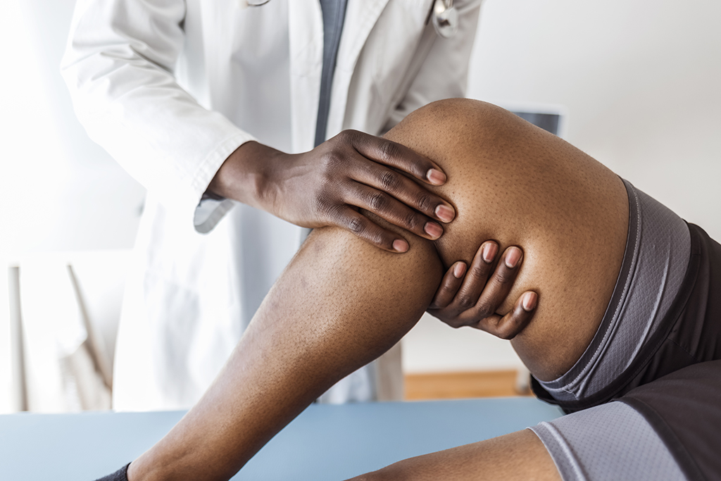
Arthritis: Characterized by inflammation of one (or more) of the body’s joints as a result of injury, wear and tear over time, or systemic disease, arthritis typically leads to pain, swelling and stiffness in the affected areas of a patient’s body. While the condition can affect any joint in the body, it is most common in weight-bearing joints such as the knees, hips and spine. The many types of arthritis include osteoarthritis, inflammatory arthritis, septic (infectious) arthritis and posttraumatic arthritis. Over time, inflammation in arthritic joints can destroy cartilage and eventually may even cause long-lasting or even permanent disability.
There is no cure for arthritis, but a range of treatment options are available that can relieve the pain it causes, along with reducing the likelihood of the condition leading to disability. Nonsurgical treatment options include medications, exercise and therapy. Surgical treatment options include total joint replacement, partial joint replacement, realignment of the joints, removal of any diseased or damaged joint lining, and fusing the bones in the joint together to limit its motion and thereby relieve pain.
Common shoulder problems: Learn more here.
Frozen shoulder: Also called adhesive capsulitis, frozen shoulder is a relatively common condition (affecting 2% of people, according to the American Academy of Orthopaedic Surgeons) that sees the capsule surrounding the shoulder joint and rotator cuff tendons grow thicker. It often presents following extended periods of shoulder immobility, such as when the shoulder is underused due to pain after an injury. Symptoms of frozen shoulder include pain and stiffness in the shoulder, along with restricted shoulder movement. Typically, after an initial worsening of symptoms, frozen shoulder will heal on its own, with the return to normal strength and range of motion taking six months to two years. Nonsurgical treatment options for the condition include the application of heat or cold to the shoulder, medicine to reduce pain and inflammation, physical therapy, electrical stimulation of nerves and muscles in the area, and cortisone injections. When symptoms persist after conservative treatments, surgical treatment options can include arthroscopic surgery, open surgery and shoulder replacement.
Rotator cuff tear: A group of muscles and tendons surrounding the shoulder joint that help to keep the upper arm firmly in the shoulder socket, the rotator cuff also assists with arm movement, playing a major role in arm lifting and rotation. And according to the American Academy of Orthopaedic Surgeons, rotator cuff tears are among the most common causes of shoulder pain and disability for U.S. adults, spurring nearly 2 million physician visits annually. Tears often begin with tendon inflammation in the rotator cuff, causing pain when attempting to raise the affected arm or lift something with it. From there, pain can grow progressively worse, with additional symptoms including shoulder weakness and a cracking sensation when attempting to move the arm into certain positions. Tears that occur suddenly, such as in a fall, can cause intense pain right away and may be accompanied by a snapping sensation and immediate upper arm weakness. Continuing to use the arm without treatment after a tear can lead to further damage, potentially making the tear larger. Treatment options for rotator cuff tears typically include rest, activity modification, nonsteroidal anti-inflammatory drugs (NSAIDs) to reduce pain and swelling, and exercise and physical therapy to restore strength and movement in the shoulder. Steroid injection may be considered if more conservative therapies prove ineffective, with surgical treatment as another option when nonsurgical treatments fail to resolve the pain.
Watch Dr. Claude Jarrett’s M.E.D. School for Patients about Rotator Cuff Injuries.
Synovitis: An inflammation of the membrane (called the synovium) that lines the moveable joints in the body such as the knees, the shoulders and the hips, synovitis is categorized into two main types — transient (or toxic) synovitis and arthritis-associated synovitis. Transient synovitis is a common, sudden-onset cause of hip pain among children, typically between the ages of 3 and 10. It can cause affected children to walk with a limp or have other problems with walking, and it usually resolves on its own within seven to 10 days. Nonsteroidal anti-inflammatory drugs (NSAIDs) may be prescribed to reduce a patient’s pain, and persistent cases may call for arthroscopy to remove inflamed tissue. Synovitis associated with arthritis is most often seen in the knees, shoulders or hips of patients with rheumatoid arthritis, juvenile arthritis, psoriatic arthritis, gout or lupus. Other, smaller joints such as those in the wrist, fingers and ankles can also be affected. Symptoms of arthritis-associated synovitis include pain, and warmth and swelling in the affected joint, and a progression of the condition can result in damage to the cartilage and bone of the joint as a result of the synovium’s inflammation. Treatment options for arthritis-associated synovitis include nonsteroidal anti-inflammatory drugs (NSAIDs) and corticosteroids.
Ankylosing spondylitis: Most often seen in the spine and back, but sometimes affecting joints elsewhere such as the hips and shoulder, ankylosing spondylitis is a chronic rheumatic disease that causes the joints to become inflamed or even fused together. When the condition affects the spine and back, it can result in a stooped posture in the patient. Further, some patients with ankylosing spondylitis can experience inflammation of the eye and/or issues with the heart valves.
Treatment options for the condition can include weight control, exercises designed to increase range of motion and strengthen muscles used for joint stabilization, and medications including non-steroidal anti-inflammatory drugs (NSAIDs), disease-modifying anti-rheumatic drugs (DMARDs), and corticosteroids.
Curvature of the spine: When viewed from the side, a natural curvature to the spine is normal in all humans. But when an X-ray is taken directly from the back, the spine should go straight up and down, without any noticeable curvature to the left or right. And when such an X-ray shows a curvature that looks similar to the letter “C” or the letter “S,” this can be problematic for the patient. This is often the case with a number of conditions collectively called “curvature of the spine.” For more specific information on some of these conditions, see scoliosis and kyphosis.
Degenerative disc disease: Also known as DDD and typically associated with advanced age, degenerative disc disease is characterized by a shrinking of the discs in the spine as they dry out and weaken over time. This typically causes the spaces between the vertebrae to tighten, leading to pain as a result of compressed nerve roots. Further, if the discs deteriorate to the point where they collapse, this can result in the facet joints in the spine rubbing together, causing pain and stiffness in the patient, and potentially leading to osteoarthritis. Additional symptoms of degenerative disc disease can include sciatica, pain that ebbs and flows or gets worse when standing, walking or sitting, trouble lifting and bending, and pain down the hip and buttock. Nonsurgical treatments for degenerative disc disease can include exercise, application of heat and/or cold packs, medication, spinal injections, and alternative treatments such as massage and physical therapy. When other treatment options prove ineffective and the pain is chronic, surgical treatment may be considered, with options including discectomy, microdiscectomy, disc replacement, spinal fusion, laminectomy, laminotomy and laminoplasty.
Discectomy and microdiscectomy: Performed in an effort to remove part of a herniated disc and relieve the pressure it is putting on a nerve to cause either back or leg pain, these surgical procedures typically deliver high levels of success — with success rates as high as 90% when most of the pain is in the leg, according to the American Academy of Orthopaedic Surgeons. By using a special instrument to view the problem disc and affected nerves, a microdiscectomy can allow for the procedure to be performed using smaller incisions. In some cases, the damaged disc can be removed altogether and replaced with an artificial one to help restore the natural distance between a pair of vertebrae.
Herniated disc: Most often seen in the lower back, a herniated disc — sometimes referred to as a bulging, protruding, slipped or ruptured disc — is among the most common causes of lower back pain and leg pain (or sciatica) in patients. The condition, often caused by wear and tear or a sudden injury, sees the soft, jelly-like center of the discs that provide cushioning between the vertebrae in the spine begin bulging into the spinal canal, putting pressure on the spinal nerves. When a disc herniates in the lower back, symptoms often include leg pain (typically below the knee), weakness, numbness, tingling in a leg or buttock, and, in extreme cases, loss of bladder or bowel control. Herniations in the upper back, on the other hand, can lead to burning pain in the neck and shoulder (and sometimes down the arm), headaches, and weakness, tingling and/or numbness in one arm. Nonsurgical treatments for a herniated disc can include exercise, medication, hot and/or cold packs, spinal injections, and massage. Surgery may be recommended in chronic cases, with options here including discectomy or microdiscectomy, spinal fusion, laminectomy, laminotomy, laminoplasty, and disc replacement.
Kyphosis: When viewed from the side, a natural curvature to the spine is normal in all humans. But in patients with kyphosis, a spinal disorder also referred to as roundback, the spine’s natural curvature is exaggerated, causing the back to look especially rounded or hunched. Sometimes caused as a result of slouching and poor posture, which can stretch spinal ligaments and lead to an increased spinal curvature, kyphosis of this kind is rarely painful and can sometimes be corrected with strengthening exercises. Congenital kyphosis, on the other hand, which sometimes occurs when an infant’s spinal column hasn’t properly developed, is likely to worsen as the child grows, often requiring surgery to correct.
Laminectomy: Often performed in conjunction with discectomy and/or fusion as part of a decompression surgery, a laminectomy is a surgical procedure focused on the complete removal of the lamina, the small bones found on the backside of a vertebra, in an effort to access the spinal cord or relieve pressure on the spinal nerves.
Laminotomy: A laminotomy is a surgical procedure to remove part of the lamina, the small bones found on the backside of a vertebra, in an effort to alleviate pressure on the nerves of the spine.
Paget’s disease of bone: A chronic disease affecting the skeleton, Paget’s disease of bone is a condition that causes the development of enlarged, deformed bones in the patients who have it. It is characterized by an excessive breakdown and formation of bone tissue (via a process called “remodeling”) — possibly caused by a viral infection along with hereditary factors — that causes a weakening of the bones. Noticeable symptoms don’t always present, but when they do, they can include bone pain, headaches, hearing loss, pressure on the nerves, increased head size, bowing of limb(s), curvature of the spine and cartilage damage, with the ones appearing largely dependent on the specific bones affected. Treatment options can include nonsteroidal anti-inflammatory drugs (NSAIDs) to relieve pain, calcitonin injection and/or drugs called bisphosphonates to help reduce pain and hinder disease progression, and exercise to help maintain healthy bones, control weight and keep joints moving. In some extreme cases, surgery may be required for purposes such as repairing a fracture or relieving pressure on nerves in the spine or skull.
Ruptured disc: See herniated disc.
Scheuermann’s disease: See curvature of the spine.
Sciatica: A condition characterized by pain and numbness in the lower back, down through the buttocks and down one leg, sciatica is caused by compression of the sciatic nerve, a large nerve that runs from the lower end of the spinal column down to the bottom of the leg. The back pain sciatica causes typically delivers shock-like or burning sensations, and additional symptoms can include loss of motor control in the affected leg. The sciatic nerve compression behind the condition can be caused by a ruptured disc, degeneration of the sciatic nerve root, or tumors or cysts in the lower spine. With ample time and rest, the condition typically resolves on its own. Nonsurgical treatment options can include nonsteroidal anti-inflammatory drugs (NSAIDs) to relieve pain, application of heat or cold to the affected muscles, stretching exercises, or cortisone-like injections. In cases that don’t resolve with nonsurgical treatment, a laminotomy or a laminectomy with discectomy may be performed to address a herniated disc or bone spurs that may be causing the problems.
Scoliosis: A condition that typically develops during childhood, scoliosis is marked by an abnormal curve of the spine to the side. In more severe cases, the spinal curvature may cause one of the patient’s shoulders to be lower than the other and/or may cause the ribs or hips to protrude more on one side. While cosmetics can be a considerable concern for many scoliosis patients, the curve of the spine is typically slight, and back pain is rarely a problem for most patients. The condition can progress if left untreated, though, and breathing difficulties can present if the spinal curvature is especially severe. Treatment options for scoliosis can include observation to keep an eye on the extent of the spinal curvature, bracing to enable routine activities and to prevent the spinal curvature from getting worse, and in a small percentage of cases, surgery to straighten the spine (typically performed only when the curve is greater than 50 degrees).
Slipped disc: See herniated disc.
Spinal cysts: An enclosed sac found in the spine that is often filled with fluid, a spinal cyst can accompany a spinal tumor or appear on its own. And like a spinal tumor, when a spinal cyst grows large enough, it can apply pressure to the spinal column and/or to the nerves within the spine, causing pain and other problematic symptoms such as weakness or stiffness in the back, shoulders, arms or legs, along with headaches and numbness. And when problems persist, surgical intervention is often recommended.
Spinal fusion: Spinal fusion is a surgical procedure that sees two or more spinal vertebrae fused together using metal rods and/or a bone graft in an effort to limit pain-causing motion in the spine and/or to restore spinal stability. More often performed on the lower back than the upper back, the procedure is typically employed to relieve serious symptoms of back problems including fractured vertebra(e), herniated discs, spinal stenosis and scoliosis. It can be performed using an anterior (from the front) or posterior (from the back) approach. Minimally invasive techniques have also been developed, in some cases enabling the procedure to be performed using smaller incisions.
Spinal stenosis: A common cause of low back pain and/or leg pain, spinal stenosis is the narrowing of the passageway that the spinal cord runs through in the spine. This can cause the spinal cord and/or other nerves in the spine to be pinched, leading to pain, numbness and (in extreme cases) loss of function in the body. Most commonly seen in adults over age 50, spinal stenosis typically results from the gradual wear-and-tear of the spinal canal seen with aging — but it can also be caused by injury or present at birth in patients born with an unusually narrow spinal canal. Symptoms of spinal stenosis can include back pain, a burning sensation in the buttocks and/or legs, numbness and/or tingling in the buttocks and/or legs, and leg weakness. Nonsurgical treatment options include anti-inflammatory medications, physical therapy, lumbar traction, chiropractic manipulation and steroid injections. Surgical options including laminectomy and spinal fusion may be considered in more extreme cases.
Spinal tumors: Classified as either benign (non-cancerous) or malignant (already or becoming cancerous), spinal tumors can present problems in the spinal canal either way by creating pressure on the canal or the nerves within. Growth of a spinal tumor can also lead to bone loss or bone displacement in the vertebral column. Pain, weakness, sensory issues and motor problems are among the symptoms spinal tumors can cause, and surgical intervention is often recommended to facilitate their removal.
Spondylolysis and spondylolisthesis: Spondylolysis and spondylolisthesis are relatively common causes of pain in the lower back for children and adolescents. Spondylolysis is characterized by weakness or a stress fracture in a vertebrae of the lower spine. This can be caused by an accident or a degenerative condition, or it can be present at birth. When the stress fracture weakens the bone to such an extent that the affected vertebra shifts or slips out of place, this is called spondylolisthesis. Many cases of spondylolysis and spondylolisthesis see no symptoms present and are only discovered when X-rays are used to diagnose other conditions. When symptoms do present in patients, they often include lower back pain that can feel like muscle strain, which can radiate into the buttocks and back of the thighs and which tends to worsen with activity. In patients with spondylolisthesis, muscle spasms caused by the condition can lead to back stiffness, hamstring tightness and problems with standing or walking. Extreme cases can bring tingling, numbness and/or weakness in one or both of the legs. Nonsurgical treatment options aimed at reducing pain and allowing a recent fracture to heal include rest, nonsteroidal anti-inflammatory drugs (NSAIDs), physical therapy and bracing. In severe cases, surgical options including spinal fusion and (if needed) a procedure to relieve pressure on the spinal nerve roots may be considered.
Strains: A strain is an injury to a muscle or tendon (the fibrous tissue that connects muscles to bones) in the body. A strain can be characterized by a pull, stretch or tear of a muscle or tendon, which often occurs in the back, leg or elbow. Regularly seen strain symptoms can include pain, swelling, inflammation, muscle spasms, muscle weakness and cramping. Initial treatment for strains include the RICE method and exercises to relieve pain and restore lost mobility. In extreme cases of muscle or tendon tearing, surgical treatment is sometimes considered.
Arthritis: Characterized by inflammation of one (or more) of the body’s joints as a result of injury, wear and tear over time, or systemic disease, arthritis typically leads to pain, swelling and stiffness in the affected areas of a patient’s body. While the condition can affect any joint in the body, it is most common in weight-bearing joints such as the knees, hips and spine. The many types of arthritis include osteoarthritis, inflammatory arthritis, septic (infectious) arthritis and posttraumatic arthritis. Over time, inflammation in arthritic joints can destroy cartilage and eventually may even cause long-lasting or even permanent disability.
There is no cure for arthritis, but a range of treatment options are available that can relieve the pain it causes, along with reducing the likelihood of the condition leading to disability. Nonsurgical treatment options include medications, exercise and therapy. Surgical treatment options include total joint replacement, partial joint replacement, realignment of the joints, removal of any diseased or damaged joint lining, and fusing the bones in the joint together to limit its motion and thereby relieve pain.
Common problems of the elbow: Learn more here.
Osteochondritis dissecans: Most commonly appearing in children and adolescents, osteochondritis dissecans is a condition caused by a loss of blood supply to a portion of bone beneath the surface of a joint — most often the knee, but sometimes affecting the ankle, elbow or other joints. This leads to a cracking and loosening of the affected portions of bone and the cartilage cushioning it, with the bone sometimes detaching completely. The most common initial symptoms are pain, swelling and tenderness in the affected joint, with more extreme cases resulting in joint weakness, sharp pain, decreased range of motion, and popping and/or locking of the joint if the bone fragment becomes lodged between bones during movement. In many cases, especially those in which the affected bone and cartilage stay in place and the patient is young and still growing, the affected joint can heal on its own. But in more extreme cases, surgery may be required.
Strains: A strain is an injury to a muscle or tendon (the fibrous tissue that connects muscles to bones) in the body. A strain can be characterized by a pull, stretch or tear of a muscle or tendon, which often occurs in the back, leg or elbow. Regularly seen strain symptoms can include pain, swelling, inflammation, muscle spasms, muscle weakness and cramping. Initial treatment for strains include the RICE (rest, ice, gentle compression and elevation) method and exercises to relieve pain and restore lost mobility. In extreme cases of muscle or tendon tearing, surgical treatment is sometimes considered.
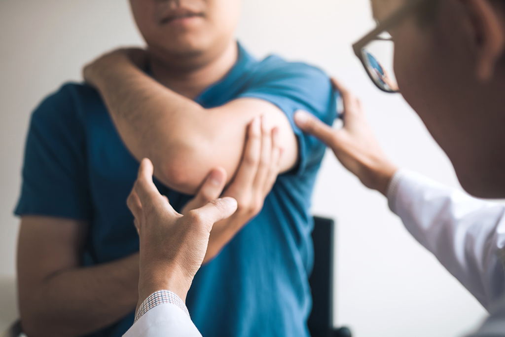
Tennis elbow: An injury that can happen to anyone who places significant stress on the elbow (and dubbed “tennis elbow” because it is seen regularly in tennis players), tennis elbow (or lateral epicondylitis) is a condition caused by elbow overuse and characterized by an inflammation of (or very small tears in) the tendons that connect the forearm muscles on the backside of the elbow. Symptoms include pain or burning on the elbow’s outer side, along with a reduced grip strength, each of which can be worsened when the forearm sees significant use. Nonsurgical treatment options, which resolve 80% to 95% of cases, include rest, medications to reduce the pain and swelling, physical therapy, bracing and steroid injections, among others. If these are ineffective, arthroscopic or open surgery may be employed to remove any diseased muscle and reattach healthy muscle tissue back to the bone.
Arthritis: Characterized by inflammation of one (or more) of the body’s joints as a result of injury, wear and tear over time, or systemic disease, arthritis typically leads to pain, swelling and stiffness in the affected areas of a patient’s body. While the condition can affect any joint in the body, it is most common in weight-bearing joints such as the knees, hips and spine. The many types of arthritis include osteoarthritis, inflammatory arthritis, septic (infectious) arthritis and posttraumatic arthritis. Over time, inflammation in arthritic joints can destroy cartilage and eventually may even cause long-lasting or even permanent disability.
There is no cure for arthritis, but a range of treatment options are available that can relieve the pain it causes, along with reducing the likelihood of the condition leading to disability. Nonsurgical treatment options include medications, exercise and therapy. Surgical treatment options include total joint replacement, partial joint replacement, realignment of the joints, removal of any diseased or damaged joint lining, and fusing the bones in the joint together to limit its motion and thereby relieve pain.
Carpal tunnel syndrome: A condition that affects the hands, wrists and forearms, carpal tunnel syndrome (CTS) is characterized by numbness, tingling, weakness and pain in the upper extremities caused by a squeezing or compression of the median nerve at the wrist. CTS is considered a repetitive stress injury, as it is often caused over time by repetitive motions, often associated with job functions. These can include typing, recurrent use of tools requiring the same hand/arm movement over and over again, and regularly repeated arm/hand motions such as turning or grasping. Heredity, pregnancy, and health conditions such as diabetes and rheumatoid arthritis are also considered risk factors for CTS.
Early in the condition’s progression, symptoms can often be relieved by taking simple steps such as avoiding activities that aggravate the condition, wearing a wrist splint during sleep, doing exercises that keep the median nerve mobile and administering a steroid injection into the affected area. If pressure on the median nerve is sustained, though, symptoms can grow worse and nerve damage can set in, in some cases requiring surgery to ease the pressure.
Common problems of the hands and wrists: Learn more here.
Sprains: Sprains are injuries characterized by tears and/or stretches of the ligaments, the tough, fibrous bands of tissue in the body that connect the ends of bones together and serve to stabilize and support the body’s joints. The most commonly sprained areas of the body are the ankles, knees and wrists. Common symptoms of sprains include pain, inflammation, bruising and swelling, with severe cases/complete ligament tears leading to significant instability. Initial treatment for sprains include the RICE method and physical therapy, with periods of bracing sometimes required for moderate-level sprains. In the most extreme cases, surgical treatment to repair torn ligaments is sometimes considered.
Writer’s cramp: See dystonia.
Arthritis: Characterized by inflammation of one (or more) of the body’s joints as a result of injury, wear and tear over time, or systemic disease, arthritis typically leads to pain, swelling and stiffness in the affected areas of a patient’s body. While the condition can affect any joint in the body, it is most common in weight-bearing joints such as the knees, hips and spine. The many types of arthritis include osteoarthritis, inflammatory arthritis, septic (infectious) arthritis and posttraumatic arthritis. Over time, inflammation in arthritic joints can destroy cartilage and eventually may even cause long-lasting or even permanent disability.
There is no cure for arthritis, but a range of treatment options are available that can relieve the pain it causes, along with reducing the likelihood of the condition leading to disability. Nonsurgical treatment options include medications, exercise and therapy. Surgical treatment options include total joint replacement, partial joint replacement, realignment of the joints, removal of any diseased or damaged joint lining, and fusing the bones in the joint together to limit its motion and thereby relieve pain.
Hip arthroscopy: Learn more here.
Watch Dr. Chad Fortunt’s M.E.D. School for Patients about Hip Arthroscopy.
Osteoporosis: The nation’s leading cause of hip fractures according to the National Institutes of Health, osteoporosis is a condition most often seen in women of advanced age (but regularly affecting older men, too). It is characterized by a thinning and weakening of the bones as patients grow older, increasing the likelihood of bone fractures. The leading causes of this gradual bone weakening include aging (as the body naturally removes old, damaged bone matter at a higher rate than it replenishes it with new bone matter via a process called “remodeling”), hormonal changes and a genetic predisposition, with poor nutrition, intake of certain medications, health conditions and lifestyle choices also playing contributing roles. Leading ways to reduce one’s risk of developing osteoporosis include maintaining a healthy diet, taking supplements (especially vitamin D and calcium — which plays a major role in osteoporosis prevention), exercising regularly, avoiding tobacco and limiting alcohol consumption, taking medications to lower the risk of bone fractures, and (for women) beginning estrogen replacement therapy if postmenopausal. Treatment options for osteoporosis include calcium and vitamin D supplements, weight-bearing exercises, physical therapy, and a range of prescription medications.
Synovitis: An inflammation of the membrane (called the synovium) that lines the moveable joints in the body such as the knees, the shoulders and the hips, synovitis is categorized into two main types — transient (or toxic) synovitis and arthritis-associated synovitis. Transient synovitis is a common, sudden-onset cause of hip pain among children, typically between the ages of 3 and 10. It can cause affected children to walk with a limp or have other problems with walking, and it usually resolves on its own within seven to 10 days. Nonsteroidal anti-inflammatory drugs (NSAIDs) may be prescribed to reduce a patient’s pain, and persistent cases may call for arthroscopy to remove inflamed tissue. Synovitis associated with arthritis is most often seen in the knees, shoulders or hips of patients with rheumatoid arthritis, juvenile arthritis, psoriatic arthritis, gout or lupus. Other, smaller joints such as those in the wrist, fingers and ankles can also be affected. Symptoms of arthritis-associated synovitis include pain, and warmth and swelling in the affected joint, and a progression of the condition can result in damage to the cartilage and bone of the joint as a result of the synovium’s inflammation. Treatment options for arthritis-associated synovitis include nonsteroidal anti-inflammatory drugs (NSAIDs) and corticosteroids.
Anterior cruciate ligament (ACL) injury: The anterior cruciate ligament, commonly called the “ACL,” is a tough, fibrous tissue in the knee that plays a critical role in preventing the shinbone from sliding beneath the thigh bone. ACL injuries are especially common among skiers, basketball players, football players and other athletes in high-demand sports, especially those who wear cleated footwear. According to the American Academy of Orthopaedic Surgeons (AAOS), ACLs are often torn or otherwise injured as a result of:
– quick twists or changes in direction
– direct impact (such as during a tackle in football)
– a reduction in speed while running
– an awkward landing in the wake of a jump
Patients who suffer ACL injuries don’t always immediately realize that they have hurt themselves. Often, a popping noise in the knee followed by the knee giving out is the first indication a patient may have of an ACL injury. This is often followed within two to 12 hours by swelling and pain in the affected knee. When an ACL injury occurs, the AAOS strongly recommends avoiding walking and/or running, as this can result in damage to the cartilage in the knee.
Other symptoms of an ACL injury can include:
– A reduction in range of motion
– Joint-line tenderness
– Tenderness when walking
Arthritis: Characterized by inflammation of one (or more) of the body’s joints as a result of injury, wear and tear over time, or systemic disease, arthritis typically leads to pain, swelling and stiffness in the affected areas of a patient’s body. While the condition can affect any joint in the body, it is most common in weight-bearing joints such as the knees, hips and spine. The many types of arthritis include osteoarthritis, inflammatory arthritis, septic (infectious) arthritis and posttraumatic arthritis. Over time, inflammation in arthritic joints can destroy cartilage and eventually may even cause long-lasting or even permanent disability.
There is no cure for arthritis, but a range of treatment options are available that can relieve the pain it causes, along with reducing the likelihood of the condition leading to disability. Nonsurgical treatment options include medications, exercise and therapy. Surgical treatment options include total joint replacement, partial joint replacement, realignment of the joints, removal of any diseased or damaged joint lining, and fusing the bones in the joint together to limit its motion and thereby relieve pain.
Changes associated with knee arthritis: Learn more here.
Chondromalacia patella: Also known as “runners knee,” this disorder is characterized by the softening of the cartilage underneath the kneecap. Typically caused by knee overuse, trauma or weakness, or a knee-structure misalignment, the condition often causes pain and swelling as a result of the kneecap rubbing against the thighbone, and it can be especially problematic when walking down stairs or downhill. Common treatment options include exercises to strengthen the affected muscles, electrical stimulation and surgery.
Knee anatomy: Learn more here.
Lateral collateral ligament (LCL) injury: A relatively rare injury according to the American Academy of Orthopaedic Surgeons, an injury to the lateral collateral ligament is typically caused by pressure or impact that pushes the knee joint from its inner side, resulting in stress to the outside of the knee joint. The lateral collateral ligament is the ligamnet in the knee that connects the thighbone to the calf bone (fibula) and helps stabilize the knee’s outer side. An LCL tear can result in pain, swelling and bruising, and most can be resolved with conservative treatments such as rest, cold packs, compression, elevation and anti-inflammatory medications. In more severe cases, surgery may be recommended.
Medial collateral ligament (MCL) injury: Most often caused by pressure or impact to the outer side of the knee, a medial collateral ligament (MCL) injury is generally a result of the over-stretching of the ligament that attaches the thighbone to the shinbone on the inside of the knee. Typically injured as a result of participation in high-contact sports, this ligament’s primary purpose is to help keep the knee’s inner side stable. Symptoms of an MCL injury often include a popping and/or sideways buckling of the knee, along with instability, pain on the inside of the knee and swelling. Many less-severe MCL injuries can be resolved with conservative treatments such as rest, cold packs, compression, elevation and anti-inflammatory medications. In more severe cases, surgery to place a hinged brace in the knee may be recommended.
Non-surgical alternatives to knee surgery: Learn more here.
Osgood-Schlatter disease: A common cause of knee pain in growing children, Osgood-Schlatter disease is characterized by a swelling of the area just below the kneecap, where the patellar tendon attaches to the shinbone. It most often develops during growth spurts in active boys ages 10 to 15, and it can lead to symptoms including a pain below the knee that is aggravated by activity, muscle tightening in the front or back of the thigh, and a painful, bony bump just beneath the kneecap. While treatment options include stretching and strengthening exercises, icing, and nonsteroidal anti-inflammatory drugs (NSAIDs), the condition will typically resolve on its own even without any treatment.
Osteochondritis dissecans: Most commonly appearing in children and adolescents, osteochondritis dissecans is a condition caused by a loss of blood supply to a portion of bone beneath the surface of a joint — most often the knee, but sometimes affecting the ankle, elbow or other joints. This leads to a cracking and loosening of the affected portions of bone and the cartilage cushioning it, with the bone sometimes detaching completely. The most common initial symptoms are pain, swelling and tenderness in the affected joint, with more extreme cases resulting in joint weakness, sharp pain, decreased range of motion, and popping and/or locking of the joint if the bone fragment becomes lodged between bones during movement. In many cases, especially those in which the affected bone and cartilage stay in place and the patient is young and still growing, the affected joint can heal on its own. But in more extreme cases, surgery may be required.
Posterior cruciate ligament (PCL) injury: Found in the knee just behind the anterior cruciate ligament (ACL), the posterior cruciate ligament (PCL) is one of a set of ligaments that connect the thighbone (femur) to the shinbone (tibia). Caused when a powerful force is exerted on a bent knee — such as a bent knee hitting a dashboard in a car crash, an athlete falling to the ground on a bent knee, or a severe twisting or contact injury during an athletic competition — an injury to the PCL can cause the shinbone to sag backward in relation to the thighbone, reducing the knee’s stability. It can also cause the ends of the shinbone and thighbone to rub directly together, weakening cartilage and potentially leading to arthritis. Symptoms of a PCL injury are often subtle and can include a popping in the knee and a buckling of the leg when weight is placed on it. Other symptoms can include pain and swelling, difficulty walking, and a feeling that the knee may “give out.” Treatment options include the RICE method (rest, ice, gentle compression and elevation) shortly after injury, immobilization and physical therapy. Surgical treatment to rebuild the ligament is sometimes recommended, especially when the PCL is injured along with other ligaments in the knee.
Runner’s knee: Also referred to as jumper’s knee and patellofemoral pain syndrome (PFPS), runner’s knee is a term used to describe pain felt in the front of the knee around the kneecap. Common in athletes, the condition is often caused by knee overuse or kneecap misalignment and can lead to pain and stiffness that can make it a challenge to perform activities such as climbing stairs, kneeling down and other activities requiring deep knee bending. Additional symptoms can include a popping or crackling sound when bending the knee or standing up after extended periods of sitting. Conservative treatments such as lowering activity levels, participating in therapeutic exercise programs, taking nonsteroidal anti-inflammatory drugs (NSAIDs) to reduce pain, using orthotics such as shoe inserts and employing the RICE method (rest, ice, compression, elevation) can often resolve symptoms. Though rare, surgical treatments such as arthroscopy and kneecap realignment may be considered.
Sprains: Sprains are injuries characterized by tears and/or stretches of the ligaments, the tough, fibrous bands of tissue in the body that connect the ends of bones together and serve to stabilize and support the body’s joints. The most commonly sprained areas of the body are the ankles, knees and wrists. Common symptoms of sprains include pain, inflammation, bruising and swelling, with severe cases/complete ligament tears leading to significant instability. Initial treatment for sprains include the RICE method and physical therapy, with periods of bracing sometimes required for moderate-level sprains. In the most extreme cases, surgical treatment to repair torn ligaments is sometimes considered.
Synovitis: An inflammation of the membrane (called the synovium) that lines the moveable joints in the body such as the knees, the shoulders and the hips, synovitis is categorized into two main types — transient (or toxic) synovitis and arthritis-associated synovitis. Transient synovitis is a common, sudden-onset cause of hip pain among children, typically between the ages of 3 and 10. It can cause affected children to walk with a limp or have other problems with walking, and it usually resolves on its own within seven to 10 days. Nonsteroidal anti-inflammatory drugs (NSAIDs) may be prescribed to reduce a patient’s pain, and persistent cases may call for arthroscopy to remove inflamed tissue. Synovitis associated with arthritis is most often seen in the knees, shoulders or hips of patients with rheumatoid arthritis, juvenile arthritis, psoriatic arthritis, gout or lupus. Other, smaller joints such as those in the wrist, fingers and ankles can also be affected. Symptoms of arthritis-associated synovitis include pain, and warmth and swelling in the affected joint, and a progression of the condition can result in damage to the cartilage and bone of the joint as a result of the synovium’s inflammation. Treatment options for arthritis-associated synovitis include nonsteroidal anti-inflammatory drugs (NSAIDs) and corticosteroids.
Arthritis: Characterized by inflammation of one (or more) of the body’s joints as a result of injury, wear and tear over time, or systemic disease, arthritis typically leads to pain, swelling and stiffness in the affected areas of a patient’s body. While the condition can affect any joint in the body, it is most common in weight-bearing joints such as the knees, hips and spine. The many types of arthritis include osteoarthritis, inflammatory arthritis, septic (infectious) arthritis and posttraumatic arthritis. Over time, inflammation in arthritic joints can destroy cartilage and eventually may even cause long-lasting or even permanent disability.
There is no cure for arthritis, but a range of treatment options are available that can relieve the pain it causes, along with reducing the likelihood of the condition leading to disability. Nonsurgical treatment options include medications, exercise and therapy. Surgical treatment options include total joint replacement, partial joint replacement, realignment of the joints, removal of any diseased or damaged joint lining, and fusing the bones in the joint together to limit its motion and thereby relieve pain.
Osteochondritis dissecans: Most commonly appearing in children and adolescents, osteochondritis dissecans is a condition caused by a loss of blood supply to a portion of bone beneath the surface of a joint — most often the knee, but sometimes affecting the ankle, elbow or other joints. This leads to a cracking and loosening of the affected portions of bone and the cartilage cushioning it, with the bone sometimes detaching completely. The most common initial symptoms are pain, swelling and tenderness in the affected joint, with more extreme cases resulting in joint weakness, sharp pain, decreased range of motion, and popping and/or locking of the joint if the bone fragment becomes lodged between bones during movement. In many cases, especially those in which the affected bone and cartilage stay in place and the patient is young and still growing, the affected joint can heal on its own. But in more extreme cases, surgery may be required.
Sprains: Sprains are injuries characterized by tears and/or stretches of the ligaments, the tough, fibrous bands of tissue in the body that connect the ends of bones together and serve to stabilize and support the body’s joints. The most commonly sprained areas of the body are the ankles, knees and wrists. Common symptoms of sprains include pain, inflammation, bruising and swelling, with severe cases/complete ligament tears leading to significant instability. Initial treatment for sprains include the RICE method and physical therapy, with periods of bracing sometimes required for moderate-level sprains. In the most extreme cases, surgical treatment to repair torn ligaments is sometimes considered.
Bunions: A bunion is a bony bump on the inside of the foot that develops when the joints of the big toe no longer fit together as they should. This often leads to pain at the spot of the bump due to irritation from footwear, as well as pain in the other toes due to overcrowding and a change in the mechanical forces exerted in the ball of the foot. Bunions can also lead to arthritis over time. Bunion treatment options can include wide-cut footwear, special padding, physical therapy and orthotic devices, as well as anti-inflammatory drugs and cortisone injections. In extreme cases, surgery can be called for to relieve the pressure caused by the bunion and repair the affected toe joint.
Clubfoot: Clubfoot, one of the most common birth defects according to the March of Dimes, is a deformity of the foot and/or ankle that causes the bottom of the foot to face inward or even upward rather than the normal downward. The condition can be seen in a single foot or both feet in affected patients, and the bones, ligaments and joints of affected extremities can all be impacted. While the condition does not cause pain in infants born with it, it can become especially problematic when the patients begin to walk. This is because it forces an affected patient to walk on the side of his or her foot rather than the sole, leading to calluses, problems wearing shoes and often-severe activity limitations. (If not treated, it can also greatly hinder the ability to walk.) Non-surgical treatment options — which see high rates of success, especially when instituted early — include stretching, casting and bracing to correct the deformity, with surgical options a possibility if these are unsuccessful.
Hammer toe: A condition marked by a deformity of the foot’s second, third or fourth toe, hammer toe is caused by a shortening of the tendons used to move the toes. The affected toe is left bent at the middle joint, making the toe resemble a hammer. A hammer toe can be painful, especially when the affected patient moves or wears shoes, and additional symptoms such as swelling or redness, difficulty walking, trouble balancing, an inability to straighten the bent toe, and corns/calluses on the top of the bent toe can present. Wearing shoes and socks with ample toe room can help resolve hammer toe, as can exercises (often guided by a physical therapist) to stretch and strengthen the affected muscles. In extreme cases, surgical treatment such as tendon transfer, tendon lengthening or joint fusion may be necessary.
Plantar fasciitis: A common cause of pain in the heel, plantar fasciitis is caused by the overuse of the plantar fascia, a strong band of tendon-like tissue that extends from the heel to the toes and supports the arch of the foot, absorbing the strains we place on our feet when they bear weight. Over time and with repeated high stresses, this tissue can develop small tears that can lead to the condition, with symptoms including inflammation and irritation on the bottom of the foot near the heel — with the pain tending to increase following periods of exercise or activity, or after extended periods of rest. Nonsurgical treatment options for plantar fasciitis can include rest, stretching exercises, modification of activities to put less stress on the joints and feet, physical therapy, nonsteroidal anti-inflammatory drugs (NSAIDs) to relieve pain, night splints, and supportive footwear. Surgical treatment may be recommended if more conservative treatments prove ineffective.
Stress fractures: Commonly seen in athletes and more common in women than men, stress fractures are typically the result of overuse of a bone. They are characterized by very small cracks in the affected bone and are most often seen in the leg or foot of patients — but can also happen in other parts of the body too, including the spine. Symptoms of a stress fracture can include pain and/or swelling at the location of the fracture, along with tenderness when the fracture site is touched. Treatment of stress fractures typically involves rest, ice-pack application, and activity modification to prevent further injury. Crutches and/or casts can sometimes be used to promote healing of a stress fracture, and placement of pins, screws or metal plates via surgery may be required in extreme cases.
Sprains: Sprains are injuries characterized by tears and/or stretches of the ligaments, the tough, fibrous bands of tissue in the body that connect the ends of bones together and serve to stabilize and support the body’s joints. The most commonly sprained areas of the body are the ankles, knees and wrists. Common symptoms of sprains include pain, inflammation, bruising and swelling, with severe cases/complete ligament tears leading to significant instability. Initial treatment for sprains include the RICE method and physical therapy, with periods of bracing sometimes required for moderate-level sprains. In the most extreme cases, surgical treatment to repair torn ligaments is sometimes considered.
Strains: A strain is an injury to a muscle or tendon (the fibrous tissue that connects muscles to bones) in the body. A strain can be characterized by a pull, stretch or tear of a muscle or tendon, which often occurs in the back, leg or elbow. Regularly seen strain symptoms can include pain, swelling, inflammation, muscle spasms, muscle weakness and cramping. Initial treatment for strains include the RICE method and exercises to relieve pain and restore lost mobility. In extreme cases of muscle or tendon tearing, surgical treatment is sometimes considered.
Strains: A strain is an injury to a muscle or tendon (the fibrous tissue that connects muscles to bones) in the body. A strain can be characterized by a pull, stretch or tear of a muscle or tendon, which often occurs in the back, leg or elbow. Regularly seen strain symptoms can include pain, swelling, inflammation, muscle spasms, muscle weakness and cramping. Initial treatment for strains include the RICE method and exercises to relieve pain and restore lost mobility. In extreme cases of muscle or tendon tearing, surgical treatment is sometimes considered.
Arthroscopy: An arthroscopy is a minimally invasive surgical procedure that allows a doctor to visualize the inside of a joint using a device called an arthroscope. This tool is a specialized type of endoscope — a tubular instrument about the size of a pencil that features an attached fiber-optic camera and lights and is designed to get into small spaces in the body. By inserting an arthroscope through an incision near a joint, a physician can perform a visual examination of the joint’s interior, then make a diagnosis regarding any conditions that may be affecting the joint.
Further, by inserting small surgical instruments such as forceps or scissors through a channel in the arthroscope, physicians can use the device to treat a range of conditions found in the joint. An arthroscope can be employed to access nearly any joint in the body, and it is commonly used to diagnose and treat problems/repair injuries in the knee, shoulder, elbow, ankle, hip and wrist.
Some of the joint conditions most commonly diagnosed and treated using an arthroscope include:
– shoulder issues such as torn rotator cuffs and frozen shoulder
– knee issues such as injuries to the cartilage and ligaments
– wrist issues such as carpal tunnel syndrome
– hip issues such as problems with the cartilage or mechanical problems such as catching, locking or buckling
– ankle issues such as sprains and loose cartilage
– tendinitis
Because of their minimally invasive nature, arthroscopic surgical procedures can often be performed on an outpatient basis and typically result in lower recovery times than open surgery, along with reduced scarring and pain.
Discectomy and microdiscectomy: Performed in an effort to remove part of a herniated disc and relieve the pressure it is putting on a nerve to cause either back or leg pain, these surgical procedures typically deliver high levels of success — with success rates as high as 90% when most of the pain is in the leg, according to the American Academy of Orthopaedic Surgeons. By using a special instrument to view the problem disc and affected nerves, a microdiscectomy can allow for the procedure to be performed using smaller incisions. In some cases, the damaged disc can be removed altogether and replaced with an artificial one to help restore the natural distance between a pair of vertebrae.
Hip arthroscopy: Learn more here.
Watch Dr. Chad Fortunt’s M.E.D. School for Patients about Hip Arthroscopy.
Joint replacement: Most often performed on the hips and knees — but also performed on other joints including the ankles, wrists, fingers, shoulders and elbows — joint replacement (also known as arthroplasty) is typically undertaken in an effort to relieve a patient’s pain and/or to help restore movement that has been limited by a problem joint. Joint replacements fall into one of two overarching categories: total joint replacement and partial joint replacement. Total joint replacement sees a problem joint removed and replaced with an artificial implant, while a partial joint replacement (also referred to as joint resurfacing) sees only the diseased and/or damaged surfaces of a joint replaced with man-made parts. Called a prosthesis, a man-made joint can be made of metal, plastic, ceramic or a combination of materials, and it’s designed to replicate the movement of the joint it is replacing. When a replacement joint is put in place, it can be either cemented into place, or put in place without cement so that bone can grow into it.
Joint resurfacing: An alternative to total joint replacement, joint resurfacing (also referred to as partial joint replacement) is often recommended for younger, more active patients whose joints have been damaged by injury or arthritis, leading to pain and limitations on movement. While the hips and knees are the most commonly resurfaced joints, the shoulders and the joints at the base of the toes (the metatarsal joints) are also candidates for joint resurfacing. One advantage of joint resurfacing over total replacement is that resurfacing leaves more original bone in place than total joint replacement does, which is beneficial should another partial or total replacement procedure be needed in the future.
Kyphoplasty: A surgical procedure employed to repair an injured or collapsed vertebra, kyphoplasty sees a balloon-like device inserted into the problem vertebra to create space before a special, glue-like substance injected into the vertebra in an effort to stabilize it and relieve any pain it may be causing. Kyphoplasty can also be used to alleviate compression of a vertebra to help re-establish its height.
Laminectomy: Often performed in conjunction with discectomy and/or fusion as part of a decompression surgery, a laminectomy is a surgical procedure focused on the complete removal of the lamina, the small bones found on the backside of a vertebra, in an effort to access the spinal cord or relieve pressure on the spinal nerves.
Laminoplasty: Performed solely on the spinal bones in the neck, a laminoplasty is a surgical procedure used to add more space in the spinal canal by cutting the lamina, the small bones found on the backside of a vertebra, to create a gap there. After a gap is created during the initial part of the procedure, it is hinged and kept open using metal hardware, bone struts, sutures, or spacers and tension bands.
Laminotomy: A laminotomy is a surgical procedure to remove part of the lamina, the small bones found on the backside of a vertebra, in an effort to alleviate pressure on the nerves of the spine.
Non-surgical alternatives to knee surgery: Learn more here.
Spinal fusion: Spinal fusion is a surgical procedure that sees two or more spinal vertebrae fused together using metal rods and/or a bone graft in an effort to limit pain-causing motion in the spine and/or to restore spinal stability. More often performed on the lower back than the upper back, the procedure is typically employed to relieve serious symptoms of back problems including fractured vertebra(e), herniated discs, spinal stenosis and scoliosis. It can be performed using an anterior (from the front) or posterior (from the back) approach. Minimally invasive techniques have also been developed, in some cases enabling the procedure to be performed using smaller incisions.
Vertebroplasty: A surgical procedure employed to repair an injured or collapsed vertebra, vertebroplasty sees a special, glue-like substance injected into the problem vertebra in an effort to stabilize it and relieve any pain it may be causing.
Age-related bone loss: See osteoporosis.
Ankylosing spondylitis: Most often seen in the spine and back, but sometimes affecting joints elsewhere such as the hips and shoulder, ankylosing spondylitis is a chronic rheumatic disease that causes the joints to become inflamed or even fused together. When the condition affects the spine and back, it can result in a stooped posture in the patient. Further, some patients with ankylosing spondylitis can experience inflammation of the eye and/or issues with the heart valves.
Treatment options for the condition can include weight control, exercises designed to increase range of motion and strengthen muscles used for joint stabilization, and medications including non-steroidal anti-inflammatory drugs (NSAIDs), disease-modifying anti-rheumatic drugs (DMARDs), and corticosteroids.
Anterior cruciate ligament (ACL) injury: The anterior cruciate ligament, commonly called the “ACL,” is a tough, fibrous tissue in the knee that plays a critical role in preventing the shinbone from sliding beneath the thigh bone. ACL injuries are especially common among skiers, basketball players, football players and other athletes in high-demand sports, especially those who wear cleated footwear. According to the American Academy of Orthopaedic Surgeons (AAOS), ACLs are often torn or otherwise injured as a result of:
– quick twists or changes in direction
– direct impact (such as during a tackle in football)
– a reduction in speed while running
– an awkward landing in the wake of a jump
Patients who suffer ACL injuries don’t always immediately realize that they have hurt themselves. Often, a popping noise in the knee followed by the knee giving out is the first indication a patient may have of an ACL injury. This is often followed within two to 12 hours by swelling and pain in the affected knee. When an ACL injury occurs, the AAOS strongly recommends avoiding walking and/or running, as this can result in damage to the cartilage in the knee.
Other symptoms of an ACL injury can include:
– A reduction in range of motion
– Joint-line tenderness
– Tenderness when walking
Arthritis: Characterized by inflammation of one (or more) of the body’s joints as a result of injury, wear and tear over time, or systemic disease, arthritis typically leads to pain, swelling and stiffness in the affected areas of a patient’s body. While the condition can affect any joint in the body, it is most common in weight-bearing joints such as the knees, hips and spine. The many types of arthritis include osteoarthritis, inflammatory arthritis, septic (infectious) arthritis and posttraumatic arthritis. Over time, inflammation in arthritic joints can destroy cartilage and eventually may even cause long-lasting or even permanent disability.
There is no cure for arthritis, but a range of treatment options are available that can relieve the pain it causes, along with reducing the likelihood of the condition leading to disability. Nonsurgical treatment options include medications, exercise and therapy. Surgical treatment options include total joint replacement, partial joint replacement, realignment of the joints, removal of any diseased or damaged joint lining, and fusing the bones in the joint together to limit its motion and thereby relieve pain.
Avascular necrosis: See osteonecrosis.
Back pain: One of the most common medical problems in the nation, back pain afflicts nearly 40% of American adults, according to the National Center for Health Statistics. With symptoms ranging from muscle aches and limited flexibility to sharp pain and an inability to stand up straight, back pain can be caused by factors, including:
– advanced age
– obesity
– muscle weakness
– muscle strain
– damage to the discs that provide cushioning between the bones (vertebrae) in the spine
– poor posture
– conditions affecting the spine such as scoliosis, osteoarthritis, rheumatic disease, osteoporosis and more
While many back-pain problems can eventually lessen or even go away with rest and recovery, the American Academy of Family Physicians (AAFP) warns that any back pain that persists, extends down into the legs and/or is accompanied by numbness or tingling should be taken seriously and spur a consultation with a physician. Treatment options for back pain can range from heat or ice treatments, massages, physical therapy and medications such as muscle relaxers or cortisone injections to provide temporary relief, to surgical procedures such as disc removal or replacement, spinal fusion and laminectomy.
Bone cancer: According to the National Cancer Institute, cancer that arises in the bones or joints is relatively rare, with roughly 3,900 new cases diagnosed in the U.S. each year — representing 0.2% of all new cancer cases nationally.
Some of the most common types of primary bone cancer include:
– osteosarcoma, the most prevalent type of bone cancer, which develops in growing bone tissue and is most common in children and adolescents
– chondrosarcoma, which arises in the cartilage — most commonly in the pelvis, legs and shoulders — and is most often diagnosed in adults
– Ewing’s sarcoma, which is believed to start in immature nerve tissue in bone marrow and is most often diagnosed in children and adolescents
Symptoms of bone cancer can include severe pain, bone weakening and a limitation of normal movement, along with fatigue, fever, weight loss and anemia. Treatments for bone cancer can include surgery, chemotherapy and radiation.
Bone fractures: A bone fracture is a break in a bone typically caused by a force such as a blow, fall or other trauma, or by a condition such as a bone tumor or osteoporosis. According to the American Academy of Orthopaedic Surgeons, there are five basic types of bone fracture. These include:
– A simple fracture, which sees a bone broken in a single place and not breaking through the skin
– A compound fracture, also known as an open fracture, in which the bone breaks through the skin
– A transverse fracture, in which the break presents at a right angle to the bone’s length
– A greenstick fracture, in which the break is seen on a single side of the bone and causes the bone to bend
– A comminuted fracture, which leaves the bone in at least three fragments
Treatment of bone fractures can range from rest and immobilization via the placement of a cast to surgical placement of wires, plates, nails, rods or screws.
Bone tumors, benign: Benign bone tumors are not cancerous and, in most cases, are non-life-threatening and will not spread to other parts of the body. These noncancerous tumors are more common than malignant bone tumors, which are cancerous, can spread to other parts of the body, and can threaten the life of affected patients.
Some of the most common types of benign bone tumors include:
– Osteochondroma: Most often found in teens and young adults, solitary osteochondromas are the most common type of benign (noncancerous) bone tumors diagnosed. The condition is characterized by an abnormal, bony growth consisting of bone and cartilage that projects from a bone surface near a growth plate.
– Fibrous dysplasia: A common type of benign (noncancerous) bone tumor, fibrous dysplasia is a bone condition that sees part(s) of a bone develop abnormally, with abnormal fibrous tissue developing in place of normal bone. The abnormal tissue can grow and expand over time, leading to pain (in some patients) and weaker bones, and raising the risk of bone fractures or deformity.
– Giant cell tumors: A type of benign (noncancerous) tumor, giant cell tumors most often appear in young adults and are typically found at the ends of the body’s long bones, often appearing close to the knee joint in the lower end of the thighbone or upper end of the shinbone. Capable of causing extreme pain, giant cell tumors tend to be aggressive, and if not treated, they can grow and damage the bones where they appear.
– Unicameral bone cysts (solitary bone cysts): Also known as a solitary or simple bone cyst and most common in children and adolescents, a unicameral bone cyst is a benign (noncancerous) bone tumor characterized by a fluid-filled cavity within the bone. Most commonly found in “long bones” such as those in the upper arm and thigh, these cysts can weaken the bone, leading to an increased risk of fracture.
– Osteoid osteomas: Small, benign (noncancerous) bone tumors typically found in the body’s longer arm and leg bones, osteoid osteomas most often occur in children and young adults. Typically less than 1.5 cm in size, these tumors do not grow in size, but they can be quite painful and can cause the formation of reactive bone around them. In some cases that occur in the spine, they can also lead to scoliosis.
– Benign cartilage tumors: Most commonly found in the skeletons of children, these noncancerous lesions originate in the cartilage. Types of benign cartilage tumors include exostoses, enchondromas, periosteal chondromas, chondromyxoid fibroma and chondroblastoma.
Bunions: A bunion is a bony bump on the inside of the foot that develops when the joints of the big toe no longer fit together as they should. This often leads to pain at the spot of the bump due to irritation from footwear, as well as pain in the other toes due to overcrowding and a change in the mechanical forces exerted in the ball of the foot. Bunions can also lead to arthritis over time. Bunion treatment options can include wide-cut footwear, special padding, physical therapy and orthotic devices, as well as anti-inflammatory drugs and cortisone injections. In extreme cases, surgery can be called for to relieve the pressure caused by the bunion and repair the affected toe joint.
Bursitis: Bursitis is an inflammation of the bursa, a small, fluid-filled sack that serves as a cushion between a tendon and a bone, such as those in the shoulder, elbow, wrist, hip, knee and ankle. Caused by overuse, injury, infection, diseases such as arthritis or diabetes, or abnormal joint positioning, bursitis can lead to pain and stiffness in patients that is intensified by movement or that flares up when trying to sleep. Common treatment options include rest, splints or other supports that restrict movement, anti-inflammatory medications and/or corticosteroid injections, administration of ice or moist heat, physical therapy, and (in severe cases) surgical procedures such as arthroscopy.
Carpal tunnel syndrome: A condition that affects the hands, wrists and forearms, carpal tunnel syndrome (CTS) is characterized by numbness, tingling, weakness and pain in the upper extremities caused by a squeezing or compression of the median nerve at the wrist. CTS is considered a repetitive stress injury, as it often caused over time by repetitive motions, often associated with job functions. These can include typing, recurrent use of tools requiring the same hand/arm movement over and over again, and regularly repeated arm/hand motions such as turning or grasping. Heredity, pregnancy, and health conditions such as diabetes and rheumatoid arthritis are also considered risk factors for CTS.
Early in the condition’s progression, symptoms can often be relieved by taking simple steps such as avoiding activities that aggravate the condition, wearing a wrist splint during sleep, doing exercises that keep the median nerve mobile and administering a steroid injection into the affected area. If pressure on the median nerve is sustained, though, symptoms can grow worse and nerve damage can set in, in some cases requiring surgery to ease the pressure.
Watch Dr. Claude Jarrett’s M.E.D. School for Patients about Carpal Tunnel Syndrome.
Cervical spondylotic myelopathy (CSM): Also known as spinal cord compression, cervical spondylotic myelopathy is a condition of the neck that appears when, over time, wear and tear of the spine leads to a squeezing/compression of the spinal cord. Identified by the American Academy of Family Physicians as the leading spinal cord problem in Americans age 55 and older, CSM commonly exhibits symptoms such as neck stiffening, weakness in the extremities, a numbness in the hands, pain in the arms, and a decline in balance and coordination. Nonsurgical treatment options for CSM include use of a soft cervical collar, physical therapy, and medications such as anti-inflammatory drugs, oral corticosteroids, epidural steroid injections and, in more extreme cases, typically limited courses of narcotics. Surgical intervention to relieve the pressure on the spinal cord is also an option in some cases.
Chondromalacia patella: Also known as “runners knee,” this disorder is characterized by the softening of the cartilage underneath the kneecap. Typically caused by knee overuse, trauma or weakness, or a knee-structure misalignment, the condition often causes pain and swelling as a result of the kneecap rubbing against the thighbone, and it can be especially problematic when walking down stairs or downhill. Common treatment options include exercises to strengthen the affected muscles, electrical stimulation and surgery.
Clubfoot: Clubfoot, one of the most common birth defects according to the March of Dimes, is a deformity of the foot and/or ankle causes the bottom of the foot to face inward or even upward rather than the normal downward. The condition can be seen in a single foot or both feet in affected patients, and the bones, ligaments and joints of affected extremities can all be impacted. While the condition does not cause pain in infants born with it, it can become especially problematic when the patients begin to walk. This is because it forces an affected patient to walk on the side of his or her foot rather than the sole, leading to calluses, problems wearing shoes and often-severe activity limitations. (If not treated, it can also greatly hinder the ability to walk.) Non-surgical treatment options — which see high rates of success, especially when instituted early — include stretching, casting and bracing to correct the deformity, with surgical options a possibility if these are unsuccessful.
Curvature of the spine: When viewed from the side, a natural curvature to the spine is normal in all humans. But when an X-ray is taken directly from the back, the spine should go straight up and down, without any noticeable curvature to the left or right. And when such an X-ray shows a curvature that looks similar to the letter “C” or the letter “S,” this can be problematic for the patient. This is often the case with a number of conditions collectively called “curvature of the spine.” For more specific information on each of these conditions, see scoliosis and kyphosis.
Degenerative disc disease: Also known as DDD and typically associated with advanced age, degenerative disc disease is characterized by a shrinking of the discs in the spine as they dry out and weaken over time. This typically causes the spaces between the vertebrae to tighten, leading to pain as a result of compressed nerve roots. Further, if the discs deteriorate to the point where they collapse, this can result in the facet joints in the spine rubbing together, causing pain and stiffness in the patient, and potentially leading to osteoarthritis. Additional symptoms of degenerative disc disease can include sciatica, pain that ebbs and flows or gets worse when standing, walking or sitting, trouble lifting and bending, and pain down the hip and buttock. Nonsurgical treatments for degenerative disc disease can include exercise, application of heat and/or cold packs, medication, spinal injections, and alternative treatments such as massage and physical therapy. When other treatment options prove ineffective and the pain is chronic, surgical treatment may be considered, with options including discectomy, microdiscectomy, disc replacement, spinal fusion, laminectomy, laminotomy and laminoplasty.
Dystonia: Characterized by involuntary and sometimes painful body movements, dystonia can affect single muscles or entire groups of them. Treatable and nonfatal, dystonia can affect just one specific part of the body (focal dystonia), multiple body parts that can be adjacent (segmental dystonia) or unrelated (multifocal dystonia), an arm and leg on the same side of the body (hemidystonia), or most or all of the body (generalized dystonia).
Focal dystonias can present in a range of types, with common ones including:
– Cervical dystonia, which is the most common type of focal dystonia and affects the muscles in the neck, often causing the head to turn to one side
– Blepharospasm, which affects the eyelids, causing them to close involuntarily
– Cranial dystonia, which affects the muscles of the neck, face and head
– Oromandibular dystonia, which affects the muscles of the tongue, lips and jaw
– Writer’s cramp, which occurs only when the patient attempts to write with the hand, causing involuntary movement in the hand muscles and sometimes extending to the forearm (Similar focal dystonias affecting other body parts/specific activities include pianist’s cramp, musician’s cramp and typist’s cramp.)
While there is no cure for dystonia, treatment options can include medications to address neurotransmitter imbalances in the brain, a suspected cause of dystonia, as well as physical and/or speech therapy, botox injections, and (in extreme cases) surgery.
Fibrous dysplasia: A common type of benign (noncancerous) bone tumor, fibrous dysplasia is a bone condition that sees part(s) of a bone develop abnormally, with abnormal fibrous tissue developing in place of normal bone. The abnormal tissue can grow and expand over time, leading to pain (in some patients) and weaker bones, and raising the risk of bone fractures or deformity. In severe cases, the condition, which is most commonly seen in the ribs, skull, thighbone, shinbone, upper arm bone and pelvis, can create a need for surgical intervention to remove affected areas of bone and repair any bone fractures or deformities.
Frozen shoulder: Also called adhesive capsulitis, frozen shoulder is a relatively common condition (affecting 2% of people, according to the American Academy of Orthopaedic Surgeons) that sees the capsule surrounding the shoulder joint and rotator cuff tendons grow thicker. It often presents following extended periods of shoulder immobility, such as when the shoulder is underused due to pain after an injury. Symptoms of frozen shoulder include pain and stiffness in the shoulder, along with restricted shoulder movement. Typically, after an initial worsening of symptoms, frozen shoulder will heal on its own, with the return to normal strength and range of motion taking six months to two years. Nonsurgical treatment options for the condition include the application of heat or cold to the shoulder, medicine to reduce pain and inflammation, physical therapy, electrical stimulation of nerves and muscles in the area, and cortisone injections. When symptoms persist after conservative treatments, surgical treatment options can include arthroscopic surgery, open surgery and shoulder replacement.
Giant cell tumor: A type of benign (noncancerous) tumor, giant cell tumors most often appear in young adults and are typically found at the ends of the body’s long bones, often appearing close to the knee joint in the lower end of the thighbone or upper end of the shinbone. Capable of causing extreme pain, giant cell tumors tend to be aggressive, and if not treated, they can grow and damage the bones where they appear. Surgery aimed at tumor removal and damage prevention is the most commonly employed treatment option for giant cell tumors, with radiation, embolization and drug therapy also among the treatment possibilities.
Hammer toe: A condition marked by a deformity of the foot’s second, third or fourth toe, hammer toe is caused by a shortening of the tendons used to move the toes. The affected toe is left bent at the middle joint, making the toe resemble a hammer. A hammer toe can be painful, especially when the affected patient moves or wears shoes, and additional symptoms such as swelling or redness, difficulty walking, trouble balancing, an inability to straighten the bent toe, and corns/calluses on the top of the bent toe can present. Wearing shoes and socks with ample toe room can help resolve hammer toe, as can exercises (often guided by a physical therapist) to stretch and strengthen the affected muscles. In extreme cases, surgical treatment such as tendon transfer, tendon lengthening or joint fusion may be necessary.
Herniated disc: Most often seen in the lower back, a herniated disc — sometimes referred to as a bulging, protruding, slipped or ruptured disc — is among the most common causes of lower back pain and leg pain (or sciatica) in patients. The condition, often caused by wear and tear or a sudden injury, sees the soft, jelly-like center of the discs that provide cushioning between the vertebrae in the spine begin bulging into the spinal canal, putting pressure on the spinal nerves. When a disc herniates in the lower back, symptoms often include leg pain (typically below the knee), weakness, numbness, tingling in a leg or buttock, and, in extreme cases, loss of bladder or bowel control. Herniations in the upper back, on the other hand, can lead to burning pain in the neck and shoulder (and sometimes down the arm), headaches, and weakness, tingling and/or numbness in one arm. Nonsurgical treatments for a herniated disc can include exercise, medication, hot and/or cold packs, spinal injections, and massage. Surgery may be recommended in chronic cases, with options here including discectomy or microdiscectomy, spinal fusion, laminectomy, laminotomy, laminoplasty, and disc replacement.
Kyphosis: When viewed from the side, a natural curvature to the spine is normal in all humans. But in patients with kyphosis, a spinal disorder also referred to as roundback, the spine’s natural curvature is exaggerated, causing the back to look especially rounded or hunched. Sometimes caused as a result of slouching and poor posture, which can stretch spinal ligaments and lead to an increased spinal curvature, kyphosis of this kind is rarely painful and can sometimes be corrected with strengthening exercises. Congenital kyphosis, on the other hand, which sometimes occurs when an infant’s spinal column hasn’t properly developed, is likely to worsen as the child grows, often requiring surgery to correct.
Lateral collateral ligament (LCL) injury: A relatively rare injury according to the American Academy of Orthopaedic Surgeons, an injury to the lateral collateral ligament is typically caused by pressure or impact that pushes the knee joint from its inner side, resulting in stress to the outside of the knee joint. The lateral collateral ligament is the ligamnet in the knee that connects the thighbone to the calf bone (fibula) and helps stabilize the knee’s outer side. An LCL tear can result in pain, swelling and bruising, and most can be resolved with conservative treatments such as rest, cold packs, compression, elevation and anti-inflammatory medications. In more severe cases, surgery may be recommended.
Medial collateral ligament (MCL) injury: Most often caused by pressure or impact to the outer side of the knee, a medial collateral ligament (MCL) injury is generally a result of the over-stretching of the ligament that attaches the thighbone to the shinbone on the inside of the knee. Typically injured as a result of participation in high-contact sports, this ligament’s primary purpose is to help keep the knee’s inner side stable. Symptoms of an MCL injury often include a popping and/or sideways buckling of the knee, along with instability, pain on the inside of the knee and swelling. Many less-severe MCL injuries can be resolved with conservative treatments such as rest, cold packs, compression, elevation and anti-inflammatory medications. In more severe cases, surgery to place a hinged brace in the knee may be recommended.
Osgood-Schlatter disease: A common cause of knee pain in growing children, Osgood-Schlatter disease is characterized by a swelling of the area just below the kneecap, where the patellar tendon attaches to the shinbone. It most often develops during growth spurts in active boys ages 10 to 15, and can lead to symptoms including a pain below the knee that is aggravated by activity, muscle tightening in the front or back of the thigh, and a painful, bony bump just beneath the kneecap. While treatment options include stretching and strengthening exercises, icing, and nonsteroidal anti-inflammatory drugs (NSAIDs), the condition will typically resolve on its own even without any treatment.
Osteoarthritis: A common type of arthritis also known as “wear and tear arthritis,” osteoarthritis is a result of the gradual breaking down and wearing down of the cartilage that cushions the joints throughout the body. As a result, the bones are left without their former cushioning at these joints, allowing them to rub together and causing pain and swelling, along with a reduction in or loss of mobility at the joint. The pain and stiffness that osteoarthritis causes also tend to worsen over time, often creating difficulty in carrying out daily activities. While there is no cure for osteoarthritis, treatment options can help affected patients better control their pain and remain active. These can include lifestyle changes, anti-inflammatory and pain-reducing medications, and physical therapy, as well as surgical options such as arthroscopy, osteotomy, joint fusion and joint replacement.
Osteochondritis dissecans: Most commonly appearing in children and adolescents, osteochondritis dissecans is a condition caused by a loss of blood supply to a portion of bone beneath the surface of a joint — most often the knee, but sometimes affecting the ankle, elbow or other joints. This leads to a cracking and loosening of the affected portions of bone and the cartilage cushioning it, with the bone sometimes detaching completely. The most common initial symptoms are pain, swelling and tenderness in the affected joint, with more extreme cases resulting in joint weakness, sharp pain, decreased range of motion, and popping and/or locking of the joint if the bone fragment becomes lodged between bones during movement. In many cases, especially those in which the affected bone and cartilage stay in place and the patient is young and still growing, the affected joint can heal on its own. But in more extreme cases, surgery may be required.
Osteochondroma: Most often found in teens and young adults, solitary osteochondromas are the most common type of benign (noncancerous) bone tumors diagnosed, accounting for 35%-40% of them. The condition is characterized by an abnormal, bony growth consisting of bone and cartilage that projects from a bone surface near a growth plate — an area where cartilage is developing near the end of a child’s long bones. While a painless bump near the joint is the most common symptom of an osteochondroma, pain and swelling can occur if the osteochondroma has a stalk that breaks off. In most cases, an osteochondroma requires no treatment beyond monitoring for changes or complications.
Osteoid osteomas: Small, benign (noncancerous) bone tumors typically found in the body’s longer arm and leg bones, osteoid osteomas most often occur in children and young adults. Typically less than 1.5 cm in size, these tumors do not grow in size, but they can be quite painful and can cause the formation of reactive bone around them. In some cases that occur in the spine, they can also lead to scoliosis. While most osteoid osteomas will disappear on their own over time, some patients may need to regularly take nonsteroidal anti-inflammatory drugs (NSAIDs) to relieve the pain they can cause. In extreme cases, surgical removal may be required.
Osteomalacia: Also known as rickets when it appears in young children, osteomalacia (derived from the Latin and Greek words for “soft bones”) is a disease that weakens the bones, often making them break easily. The disorder has a range of potential causes, with a shortage of vitamin D — a vitamin that helps the body absorb calcium — among them. Kidney disease and digestive problems can be causes, as well. The most commonly reported symptoms of osteomalacia include hip and bone pain, bone fractures, and muscle weakening, along with mobility challenges. Treatment options include vitamin D, calcium or phosphate supplements, increased exposure to sunlight, brace placement to address bone irregularities, and (in severe cases) surgical intervention.
Osteonecrosis: Also known by the names avascular necrosis, aseptic necrosis and ischemic necrosis, osteonecrosis is a condition in which a portion of bone begins to die as a result of a loss of or reduction in its blood supply. Most often diagnosed in the knees and hips, the condition typically leads to pain, swelling and a reduced range of motion in affected patients. Treatment options for osteonecrosis include medications to reduce pain, along with strengthening exercises and braces to prevent injury. Surgical options for treatment include core decompression (the removal of a bone’s inner cylinder to reduce pressure and increase blood flow), osteotomy to reshape the bone and lower the stress on affected areas, and joint replacement.
Osteopenia: Often a predecessor to osteoporosis, osteopenia is a term used to describe a decrease in bone mineral density (BMD). Most common in women over age 50, osteopenia doesn’t present specific signs or symptoms, but it can be detected early with a screening test conducted to measure bone strength. If detected, lifestyle changes can be implemented to slow osteopenia, maintain adequate bone density and prevent the onset of osteoporosis.
Osteopetrosis: A birth defect that causes affected patients to have abnormally high bone density, osteopetrosis sees the body produce more new bone than it breaks down. And while the bones of osteopetrosis patients have above-average density, their strength is below normal levels, leading to more frequent bone fractures and breaks. In addition, the patient’s immune system is often weakened as a result of connected issues with the bone marrow. Symptoms of osteopetrosis can include more-frequent-than-normal bone fractures, regular infections, and blindness and/or deafness in patients. Medications and physical therapy are among the condition’s treatment options, with bone marrow transplant a treatment option sometimes considered in severe cases.
Osteoporosis: The nation’s leading cause of hip fractures according to the National Institutes of Health, osteoporosis is a condition most often seen in women of advanced age (but regularly affecting older men, too). It is characterized by a thinning and weakening of the bones as patients grow older, increasing the likelihood of bone fractures. The leading causes of this gradual bone weakening include aging (as the body naturally removes old, damaged bone matter at a higher rate than it replenishes it with new bone matter via a process called “remodeling”), hormonal changes and a genetic predisposition, with poor nutrition, intake of certain medications, health conditions and lifestyle choices also playing contributing roles. Leading ways to reduce one’s risk of developing osteoporosis include maintaining a healthy diet, taking supplements (especially vitamin D and calcium — which plays a major role in osteoporosis prevention), exercising regularly, avoiding tobacco and limiting alcohol consumption, taking medications to lower the risk of bone fractures, and (for women) beginning estrogen replacement therapy if postmenopausal. Treatment options for osteoporosis include calcium and vitamin D supplements, weight-bearing exercises, physical therapy, and a range of prescription medications.
Paget’s disease of bone: A chronic disease affecting the skeleton, Paget’s disease of bone is a condition that causes the development of enlarged, deformed bones in the patients who have it. It is characterized by an excessive breakdown and formation of bone tissue (via a process called “remodeling”) — possibly caused by a viral infection along with hereditary factors — that causes a weakening of the bones. Noticeable symptoms don’t always present, but when they do, they can include bone pain, headaches, hearing loss, pressure on the nerves, increased head size, bowing of limb(s), curvature of the spine and cartilage damage, with the ones appearing largely dependent on the specific bones affected. Treatment options can include nonsteroidal anti-inflammatory drugs (NSAIDs) to relieve pain, calcitonin injection and/or drugs called bisphosphonates to help reduce pain and hinder disease progression, and exercise to help maintain healthy bones, control weight and keep joints moving. In some extreme cases, surgery may be required for purposes such as repairing a fracture or relieving pressure on nerves in the spine or skull.
Posterior cruciate ligament (PCL) injury: Found in the knee just behind the anterior cruciate ligament (ACL), the posterior cruciate ligament (PCL) is one of a set of ligaments that connect the thighbone (femur) to the shinbone (tibia). Caused when a powerful force is exerted on a bent knee — such as a bent knee hitting a dashboard in a car crash, an athlete falling to the ground on a bent knee, or a severe twisting or contact injury during an athletic competition — an injury to the PCL can cause the shinbone to sag backward in relation to the thighbone, reducing the knee’s stability. It can also cause the ends of the shinbone and thighbone to rub directly together, weakening cartilage and potentially leading to arthritis. Symptoms of a PCL injury are often subtle and can include a popping in the knee and a buckling of the leg when weight is placed on it. Other symptoms can include pain and swelling, difficulty walking, and a feeling that the knee may “give out.” Treatment options include the RICE method (rest, ice, gentle compression and elevation) shortly after injury, immobilization and physical therapy. Surgical treatment to rebuild the ligament is sometimes recommended, especially when the PCL is injured along with other ligaments in the knee.
Plantar fasciitis: A common cause of pain in the heel, plantar fasciitis is caused by the overuse of the plantar fascia, a strong band of tendon-like tissue that extends from the heel to the toes and supports the arch of the foot, absorbing the strains we place on our feet when they bear weight. Over time and with repeated high stresses, this tissue can develop small tears that can lead to the condition, with symptoms including inflammation and irritation on the bottom of the foot near the heel — with the pain tending to increase following periods of exercise or activity, or after extended periods of rest. Nonsurgical treatment options for plantar fasciitis can include rest, stretching exercises, modification of activities to put less stress on the joints and feet, physical therapy, nonsteroidal anti-inflammatory drugs (NSAIDs) to relieve pain, night splints, and supportive footwear. Surgical treatment may be recommended if more conservative treatments prove ineffective.
Reiter’s syndrome: Also referred to as reactive arthritis, Reiter’s syndrome is a type of arthritis that sets in as a result of (or “reaction” to) an infection in another part of the body, often the intestines or urinary tract. The condition produces symptoms that can include pain, swelling, redness and a feeling of heat in the joints (often the knees, ankles and/or feet) — and, in some cases, redness and inflammation in the eyes, along with urinary tract inflammation. Treatment options for reactive arthritis can include nonsteroidal anti-inflammatory drugs (NSAIDs), corticosteroid injections, topical corticosteroids and antibiotics.
Repetitive stress injuries: As the name implies, repetitive stress injuries — sometimes referred to as repetitive motion disorders — are caused by repetitive (often on-the-job) motions such as typing, repetitive tool use, and repetitive motions such as grasping or turning. Appearing in various areas of the body and coming in a range of different types, examples of repetitive stress injuries include carpal tunnel syndrome, tendinitis and bursitis, shin splints, plantar fasciitis, tennis elbow and trigger finger.
Often causing considerable pain that can even become debilitating, repetitive stress injuries typically present initial symptoms such as tingling, numbness or pain in the affected area, sometimes accompanied by a loss of flexibility and/or strength. The symptoms often progressively worsen without treatment, sometimes even resulting in a total loss of function in the affected area(s). Treatment options can include drugs to reduce inflammation, medications to reduce pain and swelling, icing and splints, with physical therapy often recommended after pain subsides to rebuild affected muscles and prevent future injuries. Surgery may also be recommended in extreme cases.
Rotator cuff tear: A group of muscles and tendons surrounding the shoulder joint that help to keep the upper arm firmly in the shoulder socket, the rotator cuff also assists with arm movement, playing a major role in arm lifting and rotation. And according to the American Academy of Orthopaedic Surgeons, rotator cuff tears are among the most common causes of shoulder pain and disability for U.S. adults, spurring nearly 2 million physician visits annually. Tears often begin with tendon inflammation in the rotator cuff, causing pain when attempting to raise the affected arm or lift something with it. From there, pain can grow progressively worse, with additional symptoms including shoulder weakness and a cracking sensation when attempting to move the arm into certain positions. Tears that occur suddenly, such as in a fall, can cause intense pain right away and may be accompanied by a snapping sensation and immediate upper arm weakness. Continuing to use the arm without treatment after a tear can lead to further damage, potentially making the tear larger. Treatment options for rotator cuff tears typically include rest, activity modification, nonsteroidal anti-inflammatory drugs (NSAIDs) to reduce pain and swelling, and exercise and physical therapy to restore strength and movement in the shoulder. Steroid injection may be considered if more conservative therapies prove ineffective, with surgical treatment as another option when nonsurgical treatments fail to resolve the pain.
Watch Dr. Claude Jarrett’s M.E.D. School for Patients about Rotator Cuff Injuries.
Runner’s knee: Also referred to as jumper’s knee and patellofemoral pain syndrome (PFPS), runner’s knee is a term used to describe pain felt in the front of the knee around the kneecap. Common in athletes, the condition is often caused by knee overuse or kneecap misalignment and can lead to pain and stiffness that can make it a challenge to perform activities such as climbing stairs, kneeling down and other activities requiring deep knee bending. Additional symptoms can include a popping or crackling sound when bending the knee or standing up after extended periods of sitting. Conservative treatments such as lowering activity levels, participating in therapeutic exercise programs, taking nonsteroidal anti-inflammatory drugs (NSAIDs) to reduce pain, using orthotics such as shoe inserts and employing the RICE method (rest, ice, compression, elevation) can often resolve symptoms. Though rare, surgical treatments such as arthroscopy and kneecap realignment may be considered.
Ruptured disc: See herniated disc.
Scheuermann’s disease: See curvature of the spine.
Sciatica: A condition characterized by pain and numbness in the lower back, down through the buttocks and down one leg, sciatica is caused by compression of the sciatic nerve, a large nerve that runs from the lower end of the spinal column down to the bottom of the leg. The back pain sciatica causes typically delivers shock-like or burning sensations, and additional symptoms can include loss of motor control in the affected leg. The sciatic nerve compression behind the condition can be caused by a ruptured disc, degeneration of the sciatic nerve root, or tumors or cysts in the lower spine. With ample time and rest, the condition typically resolves on its own. Nonsurgical treatment options can include nonsteroidal anti-inflammatory drugs (NSAIDs) to relieve pain, application of heat or cold to the affected muscles, stretching exercises, or cortisone-like injections. In cases that don’t resolve with nonsurgical treatment, a laminotomy or a laminectomy with discectomy may be performed to address a herniated disc or bone spurs that may be causing the problems.
Scoliosis: A condition that typically develops during childhood, scoliosis is marked by an abnormal curve of the spine to the side. In more severe cases, the spinal curvature may cause one of the patient’s shoulders to be lower than the other and/or may cause the ribs or hips to protrude more on one side. While cosmetics can be a considerable concern for many scoliosis patients, the curve of the spine is typically slight, and back pain is rarely a problem for most patients. The condition can progress if left untreated, though, and breathing difficulties can present if the spinal curvature is especially severe. Treatment options for scoliosis can include observation to keep an eye on the extent of the spinal curvature, bracing to enable routine activities and to prevent the spinal curvature from getting worse, and in a small percentage of cases, surgery to straighten the spine (typically performed only when the curve is greater than 50 degrees).
Shin splints: An overuse injury common among runners and athletes, shin splints typically present after heavy physical exertion and are characterized by pain, swelling and soreness along the shinbone’s inner edge. They are caused by an inflammation of the tendons, muscles and bone tissue around the tibia, typically as a result of overtraining, ill-fitting or worn-out footwear, and extended durations of downhill running. The pain caused by shin splints can usually be relieved with simple, conservative measures such as rest, compression, ice, shoe inserts and stretching. Surgery is very rarely needed for treatment of shin splints.
Slipped disc: See herniated disc.
Spinal cysts: An enclosed sac found in the spine that is often filled with fluid, a spinal cyst can accompany a spinal tumor or appear on its own. And like a spinal tumor, when a spinal cyst grows large enough, it can apply pressure to the spinal column and/or to the nerves within the spine, causing pain and other problematic symptoms such as weakness or stiffness in the back, shoulders, arms or legs, along with headaches and numbness. And when problems persist, surgical intervention is often recommended.
Spinal stenosis: A common cause of low back pain and/or leg pain, spinal stenosis is the narrowing of the passageway that the spinal cord runs through in the spine. This can cause the spinal cord and/or other nerves in the spine to be pinched, leading to pain, numbness and (in extreme cases) loss of function in the body. Most commonly seen in adults over age 50, spinal stenosis typically results from the gradual wear-and-tear of the spinal canal seen with aging — but it can also be caused by injury or present at birth in patients born with an unusually narrow spinal canal. Symptoms of spinal stenosis can include back pain, a burning sensation in the buttocks and/or legs, numbness and/or tingling in the buttocks and/or legs, and leg weakness. Nonsurgical treatment options include anti-inflammatory medications, physical therapy, lumbar traction, chiropractic manipulation and steroid injections. Surgical options including laminectomy and spinal fusion may be considered in more extreme cases.
Spinal tumors: Classified as either benign (non-cancerous) or malignant (already or becoming cancerous), spinal tumors can present problems in the spinal canal either way by creating pressure on the canal or the nerves within. Growth of a spinal tumor can also lead to bone loss or bone displacement in the vertebral column. Pain, weakness, sensory issues and motor problems are among the symptoms spinal tumors can cause, and surgical intervention is often recommended to facilitate their removal.
Spondylolysis and spondylolisthesis: Spondylolysis and spondylolisthesis are relatively common causes of pain in the lower back for children and adolescents. Spondylolysis is characterized by weakness or a stress fracture in a vertebrae of the lower spine. This can be caused by an accident or a degenerative condition, or it can be present at birth. When the stress fracture weakens the bone to such an extent that the affected vertebra shifts or slips out of place, this is called spondylolisthesis. Many cases of spondylolysis and spondylolisthesis see no symptoms present and are only discovered when X-rays are used to diagnose other conditions. When symptoms do present in patients, they often include lower back pain that can feel like muscle strain, which can radiate into the buttocks and back of the thighs and which tends to worsen with activity. In patients with spondylolisthesis, muscle spasms caused by the condition can lead to back stiffness, hamstring tightness and problems with standing or walking. Extreme cases can bring tingling, numbness and/or weakness in one or both of the legs. Nonsurgical treatment options aimed at reducing pain and allowing a recent fracture to heal include rest, nonsteroidal anti-inflammatory drugs (NSAIDs), physical therapy and bracing. In severe cases, surgical options including spinal fusion and (if needed) a procedure to relieve pressure on the spinal nerve roots may be considered.
Sprains: Sprains are injuries characterized by tears and/or stretches of the ligaments, the tough, fibrous bands of tissue in the body that connect the ends of bones together and serve to stabilize and support the body’s joints. The most commonly sprained areas of the body are the ankles, knees and wrists. Common symptoms of sprains include pain, inflammation, bruising and swelling, with severe cases/complete ligament tears leading to significant instability. Initial treatment for sprains include the RICE method and physical therapy, with periods of bracing sometimes required for moderate-level sprains. In the most extreme cases, surgical treatment to repair torn ligaments is sometimes considered.
Strains: A strain is an injury to a muscle or tendon (the fibrous tissue that connects muscles to bones) in the body. A strain can be characterized by a pull, stretch or tear of a muscle or tendon, which often occurs in the back, leg or elbow. Regularly seen strain symptoms can include pain, swelling, inflammation, muscle spasms, muscle weakness and cramping. Initial treatment for strains include the RICE method and exercises to relieve pain and restore lost mobility. In extreme cases of muscle or tendon tearing, surgical treatment is sometimes considered.
Stress fractures: Commonly seen in athletes and more common in women than men, stress fractures are typically the result of overuse of a bone. They are characterized by very small cracks in the affected bone and are most often seen in the leg or foot of patients — but can also happen in other parts of the body too, including the spine. Symptoms of a stress fracture can include pain and/or swelling at the location of the fracture, along with tenderness when the fracture site is touched. Treatment of stress fractures typically involves rest, ice-pack application, and activity modification to prevent further injury. Crutches and/or casts can sometimes be used to promote healing of a stress fracture, and placement of pins, screws or metal plates via surgery may be required in extreme cases.
Synovitis: An inflammation of the membrane (called the synovium) that lines the moveable joints in the body such as the knees, the shoulders and the hips, synovitis is categorized into two main types — transient (or toxic) synovitis and arthritis-associated synovitis. Transient synovitis is a common, sudden-onset cause of hip pain among children, typically between the ages of 3 and 10. It can cause affected children to walk with a limp or have other problems with walking, and it usually resolves on its own within seven to 10 days. Nonsteroidal anti-inflammatory drugs (NSAIDs) may be prescribed to reduce a patient’s pain, and persistent cases may call for arthroscopy to remove inflamed tissue. Synovitis associated with arthritis is most often seen in the knees, shoulders or hips of patients with rheumatoid arthritis, juvenile arthritis, psoriatic arthritis, gout or lupus. Other, smaller joints such as those in the wrist, fingers and ankles can also be affected. Symptoms of arthritis-associated synovitis include pain, and warmth and swelling in the affected joint, and a progression of the condition can result in damage to the cartilage and bone of the joint as a result of the synovium’s inflammation. Treatment options for arthritis-associated synovitis include nonsteroidal anti-inflammatory drugs (NSAIDs) and corticosteroids.
Tendinitis: Tendinitis is the inflammation (characterized by redness, soreness and swelling) of a tendon, which is a connective tissue in the body that attached muscle to bone. Caused by overuse, injury, infection, diseases such as arthritis or diabetes, or abnormal joint positioning, tendinitis can lead to pain and stiffness in patients that is intensified by movement or that flares up when trying to sleep. Common treatment options include rest, splints or other supports that restrict movement, anti-inflammatory medications and/or corticosteroid injections, administration of ice or moist heat, physical therapy, and (in severe cases) surgical procedures such as arthroscopy.
Tennis elbow: An injury that can happen to anyone who places significant stress on the elbow (and dubbed “tennis elbow” because it is seen regularly in tennis players), tennis elbow (or lateral epicondylitis) is a condition caused by elbow overuse and characterized by an inflammation of (or very small tears in) the tendons that connect the forearm muscles on the backside of the elbow. Symptoms include pain or burning on the elbow’s outer side, along with a reduced grip strength, each of which can be worsened when the forearm sees significant use. Nonsurgical treatment options, which resolve 80% to 95% of cases, include rest, medications to reduce the pain and swelling, physical therapy, bracing and steroid injections, among others. If these are ineffective, arthroscopic or open surgery may be employed to remove any diseased muscle and reattach healthy muscle tissue back to the bone.
Unicameral bone cyst: Also known as a solitary or simple bone cyst and most common in children and adolescents, a unicameral bone cyst is a benign (noncancerous) bone tumor characterized by a fluid-filled cavity within the bone. Most commonly found in “long bones” such as those in the upper arm and thigh, these cysts can weaken the bone, leading to an increased risk of fracture. If the risk of fracture is especially high, a surgical treatment known as aspiration and injection may be called for. This procedure sees a needle inserted into the cyst to drain the fluid within (aspiration), followed by an injection of substances meant to prevent the cyst from reappearing.
Writer’s cramp: See dystonia.
In most cases involving orthopaedic issues, more conservative treatment options such as medications, exercise and other rehabilitative or alternative therapies are the first course of action. When necessary (typically in more severe cases), a physician may also recommend moving on to surgical diagnostic and treatment options, such as arthroscopy or joint replacement (partial or full).
- Arthroscopy: An arthroscopy is a minimally invasive surgical procedure that allows a doctor to visualize the inside of a joint using a device called an arthroscope. This tool is a specialized type of endoscope — a tubular instrument about the size of a pencil that features an attached fiber-optic camera and lights and is designed to get into small spaces in the body. By inserting an arthroscope through an incision near a joint, a physician can perform a visual examination of the joint’s interior, then make a diagnosis regarding any conditions that may be affecting the joint.
Further, by inserting small surgical instruments such as forceps or scissors through a channel in the arthroscope, physicians can use the device to treat a range of conditions found in the joint. An arthroscope can be employed to access nearly any joint in the body, and it is commonly used to diagnose and treat problems/repair injuries in the knee, shoulder, elbow, ankle, hip and wrist.
Some of the joint conditions most commonly diagnosed and treated using an arthroscope include:
– shoulder issues such as torn rotator cuffs and frozen shoulder
– knee issues such as injuries to the cartilage and ligaments
– wrist issues such as carpal tunnel syndrome
– hip issues such as problems with the cartilage or mechanical problems such as catching, locking or buckling
– ankle issues such as sprains and loose cartilage
– tendinitis
Because of their minimally invasive nature, arthroscopic surgical procedures can often be performed on an outpatient basis and typically result in lower recovery times than open surgery, along with reduced scarring and pain.
- DEXA scan: Typically going by a name that includes an abbreviation for “dual-energy X-ray absorptiometry,” a DEXA scan is a diagnostic tool that is often used to measure bone density and monitor its changes over time. Utilizing an instrument that emits low-energy X-rays, the machine is often used to create images of the spine, hip, total body and wrist via a painless and noninvasive exam. This exam usually takes about 20 minutes to complete and sees patients recline on a padded table while the machine moves around them to capture images.
- RICE: An initialism for “rest, ice, compression, elevation,” RICE is a type of therapy often employed in the wake of joint injuries. Resting the joint helps give it time to heal. Ice is often used to control swelling, as is compression. And elevating the joint above the level of the heart can help reduce pain, throbbing and swelling.